Dog eye infections can be quite common, with gundogs susceptible to a number of different hereditary eye conditions that owners need to be aware of.
Healthy eyes are key for any working dog, and responsible breeders will play a key role in maintaining their dogs’ eye health, working closely with veterinary eye specialists and geneticists towards this goal.
Dog eye conditions can affect any part of the eye, from the eyelids to the lens to the retina. As a result, a basic understanding of a dog’s eye anatomy is really helpful to understand the various inherited eye conditions that can affect dogs.
The Anatomy Of A Dog’s Eye
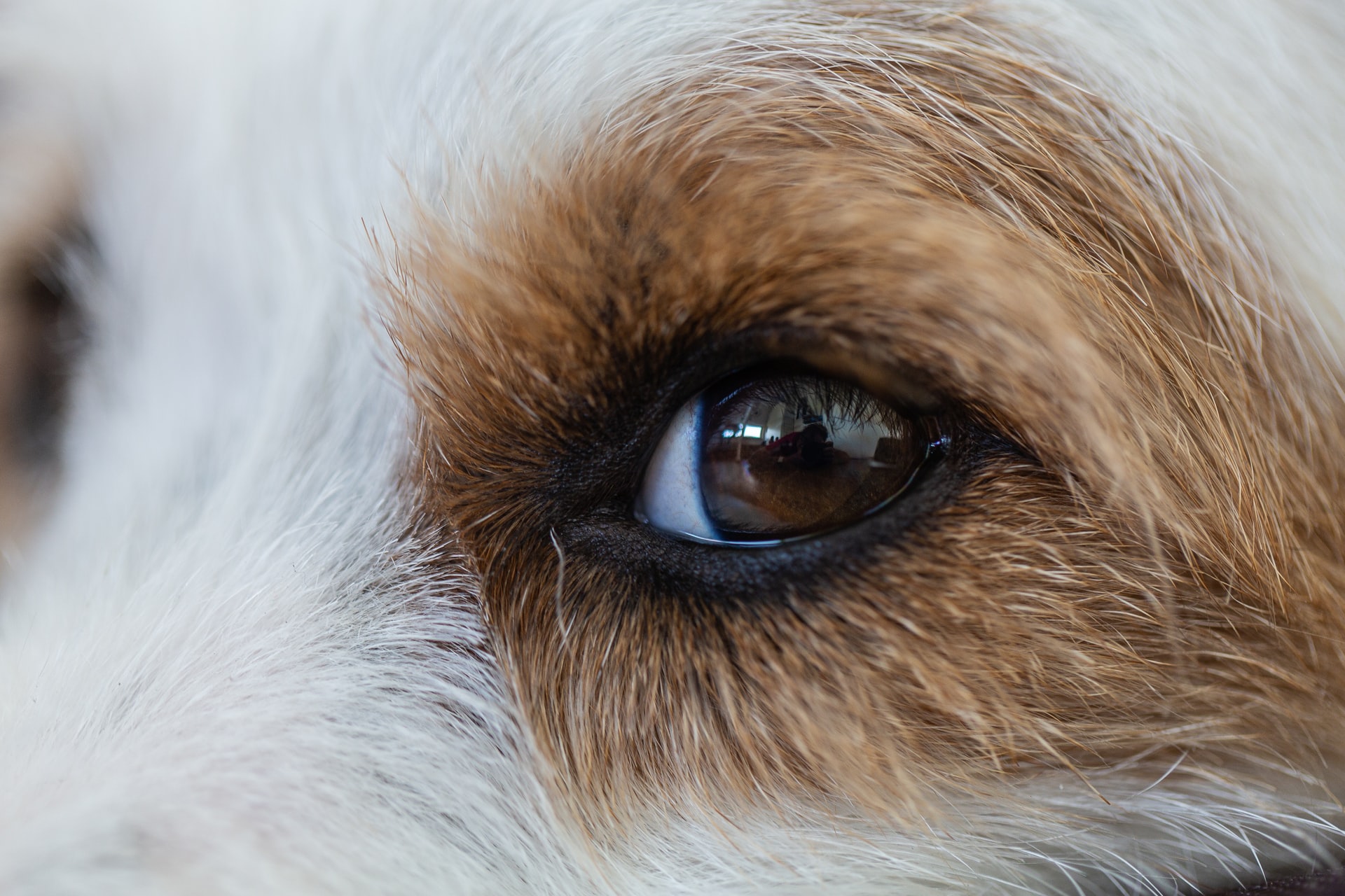
A dog’s eyeball is made up of three basic layers:
- Cornea and sclera - the outermost layer is divided into the clear window at the front of the eye called the cornea, and the white coat of the eye called the sclera. The cornea in working dogs is prone to wounding by thorns and bushes and is protected by the tear film and the eyelids. In-turning of the eyelids, called ‘entropion’, is painful and can cause scarring and ulceration
- Uvea - the middle layer of the eyeball is called the uvea and it’s this layer that forms the pupil and nourishes the inside of the eye. The lens is suspended by fine fibrils behind the pupil and focuses on the in-coming image. If the lens goes opaque, this is called a cataract - cataracts in dogs may affect vision, depending on their extent
- Retina - the innermost layer of the eye, called the retina is at the back of the eyeball and it’s this layer that forms an image and sends it to the brain via the optic nerve. Diseases where the retina stops working result in blindness with an eye that is otherwise normal in appearance
When Is A Dog Eye Test Needed?
Eye tests for dogs will always be needed. It’s important to establish that eye examination and genetic testing are never exclusive of each other and should be used in a complimentary manner.
Specifically, there are two good reasons why an eye test will never be entirely replaced by genetic testing:
- Genetic tests are not yet available for all eye conditions and eye testing is paramount in identifying potential dog eye infections and removing the affected dogs from breeding programmes
- The eye examination serves as an early warning by identifying emerging inherited eye conditions. This allows proactive breeders to prevent a disease becoming widely spread throughout the breed before it is officially acknowledged - something which is especially important in numerically small breeds
Dog Eye Test Checklist
If you’re unsure whether to get your gundog’s eyes tested for common dog eye conditions, take a look at this checklist:
1. Check Which Dog Eye Conditions Apply To Your Breed
You can find out which dog eye conditions apply to your breed via the British Veterinary Association (BVA) eye scheme web page or the Kennel Club (KC) Assured Breeders specific health requirements page.
If your breed doesn’t have a specific known inherited dog eye condition, it’s still a good idea to have a routine dog eye test carried out under the BVA/KC/ISDS eye scheme before you breed.
2. Check Which DNA Tests Your Dog Needs
Your dog may already be hereditarily clear for some DNA tests, so find this out before taking your dog for tests. For example, if both of your dog’s parents have been DNA tested as free for PRA, your dog may be hereditarily clear for this form of PRA, so an additional test is not required. This information can be found either on your KC registration document or via the KC Health Test Results Finder.
3. Is Your Dog Past The Age Of One?
Most breeders will present their dog for their first dog eye test at the age of one, just before any planned hip and elbow x-rays.
- If your dog is a breed that is prone to glaucoma, gonioscopy (the test for glaucoma in dogs) can now be carried out for the first time and does not need to be repeated for three years
- If your dog tests negative for MRD and TRD, it will be clear for life for these conditions
- Hereditary cataract and retinal degeneration can set in years later, so regular examination before each time a bitch is bred - or annually in case of a much used stud dog - is necessary to identify these conditions
- If you have dogs that are no longer active in breeding, consider having them examined when they are more than eight years old for a reduced fee. Findings here will give you valuable information if you have held back offspring of these dogs to continue your line
- If you breed litters of breeds that are known to have MRD or TRD, consider having the puppies litter screened before they leave, between 6 and 12 weeks of age
What Is The Unified Eye Testing Scheme For Dogs?
Since 1960, the BVA, the KC, and the International Sheep Dog Society have teamed to aid breeders in identifying inherited eye conditions in purebred dogs with the BVA/KC/ISDS eye scheme.
The Unified Eye Testing Scheme offers dog eye examination by a highly qualified panel of ophthalmologists throughout the UK for a set fee. This fee is less than the costs for hip and elbow scoring, as well as those for many genetic tests, and it can be reduced further by arranging group testing sessions of more than 25 dogs.
Experienced breeders will always have the eye test done first before embarking on more costly tests that might require sedation or anaesthetic. For most breeding stock, eye testing is recommended prior to each time a bitch is bred, while frequently used stud dogs should be examined annually. Tests for glaucoma in dogs are carried out on a less frequent basis - they’re currently recommended on a three-year basis.
Some breeders have avoided the Unified Eye Testing Scheme as they’re worried their name might be tarnished if one of their dogs should be affected with a hereditary condition. But any good breeder would know that if you breed long enough, you are eventually bound to encounter a problem even in the best possible line - whether it’s eyes, hips, elbows, or one of the many other genetic conditions.
What makes a good breeder is not never having a dog affected with a hereditary condition, but how they deal with it when it happens. Only this open approach can help us in understanding how conditions are passed on and in finding genetic tests that will eventually eradicate the disease.
What Happens During A Dog Eye Examination?
Here’s what to expect during a routine dog eye test:
- The dog will receive a drop of a drug that widens the pupil for a duration of approximately two hours. The drops are applied approximately 30 minutes before the eye test to make sure the pupils are well dilated
- The room in which the examination is performed is usually darkened
- A combination of a microscope and a bright illuminating lamp called a slit lamp is used to examine the front of the eye and the lens whilst the back of the eye is examined with an ophthalmoscope
- In breeds that are prone to inherited glaucoma, additional tests are carried out before the pupils are dilated
- The intraocular pressure may be measured and a small contact lens is applied to the eye after the cornea has been numbed with a drop of local anaesthetic
Is A Dog Eye Examination Painful?
None of the tests involved in a dog eye examination are painful and the most difficult thing can be to keep a wriggly patient sitting still for the duration of the test. Most gundogs are of course so well behaved that it’s a pleasure for the panellist to carry out the examination!
5 Of The Most Common Dog Eye Conditions In Gundogs
The most common inherited dog eye conditions that gundog owners needs to be aware of are:
- Cataracts in dogs
- Retinal atrophy
- Retinal pigment epithelial dystrophy
- Retinal dysplasia
- Glaucoma in dogs
Not all conditions apply to all breeds, and breeders should be aware that very few inherited dog eye conditions are congenital (present at birth). Most dog eye conditions actually set in later in life, with some dogs not affected until they are eight or 10 years old.
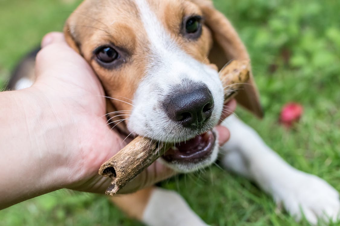
For a breeder who’s looking to establish a successful line, it’s important to not only have breeding stock examined prior to mating, but also to check their older stock for signs of late-onset inherited eye disease, as this can have important implications for the offspring they have kept.
1. Hereditary Cataracts In Dogs
Cloudy eyes in dogs can be a sign of cataracts; any opacity of the lens is described as a cataract.
Many cataracts in dogs remain small and never progress to impair vision, but others can be rapidly progressive and cause blindness even in younger dogs. Inherited forms of cataracts in dogs are usually defined foremost by their position in the lens and only secondly by their rate of progress.
Cataracts in dogs are particularly likely in the following gundog breeds:
Interestingly, these breeds show a similar clinical appearance of their inherited cataract, which manifests as an inverted, small Y-shaped opacity at the back of the lens. Some ophthalmologists refer to this type of cataract as the ‘Mercedes Benz sign’. This condition is called ‘posterior polar subcapsular cataract’, or PPC.
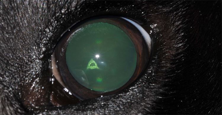
Posterior Polar Subcapsular Cataracts In Dogs
PPC is usually visible in both eyes, although occasionally only one eye is affected - or it may be apparent in one eye before the other.
In most affected dogs, cataracts in dogs appear some time during adolescence and remain small, not causing an apparent clinical problem. However, in some affected dogs, the cataract can progress and eventually cause impaired vision and blindness. This can be rectified by surgical intervention with cataract removal and lens replacement - which is of course a costly procedure that carries risks.
To date, there has been very limited progress in identifying the genetic cause of most inherited cataracts in dogs. A DNA test is not yet available for PPC in the listed breeds. An annual dog eye test remains the most efficient way to limit the impact of hereditary cataracts in dogs.
2. Generalised Progressive Retinal Atrophy (PRA) In Dogs
Light enters the retina through the clear cornea and lens; images are then formed by retinal photoreceptors, with this information sent to the area or the brain responsible for vision. Many forms of inherited retinal failure or progressive retinal atrophy (PRA) have been identified, with variations in time of onset and speed of progression between different breeds.
PRA In Irish Setters
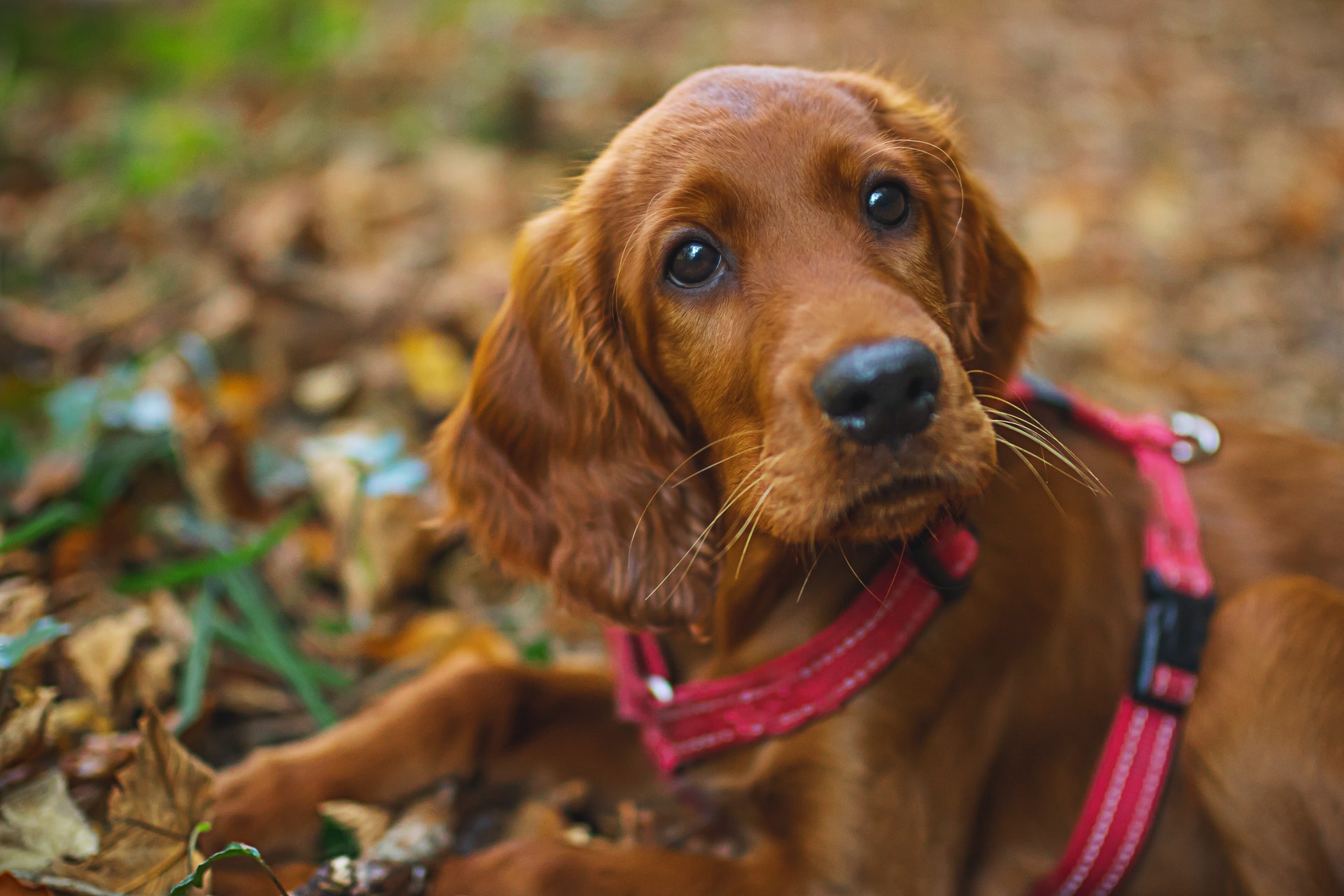
For example, Irish setters affected with PRA are born with an abnormal retina that atrophies quickly. Irish setters with PRA show signs of the disease within their first months of life and are blind by the age of one.
PRA In Cocker Spaniels
However, cocker spaniels with PRA have an initially normal retina, which then starts to fail in the adult dog. Cocker spaniels affected by retinal atrophy often don’t show signs of vision loss until late adulthood and will retain some sight until aged 10+.
PRA In Labradors
Labradors affected with PRA usually start showing visual impairment quite young - at about four years of age - and may be blind by eight or 10 years. Many labradors affected with PRA will also develop secondary cataracts.
Signs Of PRA In Dogs
Whatever the genetic variation of PRA, the clinical signs are very similar between most forms. Owners of affected dogs will usually notice impaired vision first at night-time and during times of poor illumination such as dusk and dawn. It is not uncommon to hear affected dogs collide with objects such as dustbins or cars - which are not always in the same place - when during early morning or evening walks in winter time.
As PRA progresses, the visual deficit becomes apparent during daytime and especially when dogs are exposed to unfamiliar environments. The pupils become increasingly dilated, which results in a greenish reflection similar to that of a cat’s eye at night caught in a headlamp beam. Secondary cataracts are likely to develop also.
Is There Treatment For PRA In Dogs?
Unfortunately, PRA is currently not treatable and affected dogs almost always do eventually go blind. As PRA is mostly a slowly progressive disease, affected dogs are incredibly able to adapt to their visual impairment and function to a level much better than any human, thanks to their superior ears and nose. However, those gundogs relying on sight more than their sense of smell will of course fail to perform well in the field if affected with PRA - this dog eye condition usually ends a working dog’s career.
Thanks to collaboration between veterinary ophthalmologists, geneticists and responsible breed clubs, great progress has been made in developing genetic tests for a variety of forms of PRA.
3. Retinal Pigment Epithelial Dystrophy (RPED) In Dogs
This form of retinal degeneration is now rare in the UK, but can still occasionally be seen in the spaniel breeds, as well as in labradors and golden retrievers.

RPED is associated with an abnormal Vitamin E metabolism and is diagnosed by the ophthalmologist on account of characteristic brown deposits in the retina.
The inheritance of RPED is far more complex than that of the described PRAs due to environmental factors. Clinically, affected dogs present with slowly progressive vision loss, and additional neurological deficits can be found especially in the spaniel breeds. This dog eye condition is usually identified in middle-aged dogs.
4. Retinal Dysplasia (RD) In Dogs
Retinal dysplasia (RD) describes a failure of the developing retina to unfold evenly on the nourishing tissue at the back of the eye.
Two forms of RD are distinguished in the UK:
- Total Retinal Dysplasia (TRD) - in TRD, the retina never fully attaches to the back of the eye and becomes detached in the very young pup, which usually presents blind to the breeder shortly after opening the eyes. This disease is fortunately extremely rare in the UK
- Multifocal retinal dysplasia (MRD) - this type of RD is more commonly seen in gundogs in the UK, specifically affecting the springer spaniel, the golden retriever and the labrador. In most cases, affected dogs seemingly have normal vision and only an eye examination by a veterinary ophthalmologist will find small areas where the retina is folded. However, in some springers, the condition can be more severe, affecting large areas of the retina, causing vision loss and secondary complications such as retinal detachment. A genetic test for MRD is not yet available
Both MRD and TRD are conditions that can be identified before puppies leave for their permanent homes, and the easiest time to certify puppies as ‘affected’ or ‘unaffected’ is with a litter eye screening exam.
In this examination, the entire litter, which must already be permanently identified by microchip, is presented to a panellist for examination between the ages of five and 12 weeks. Each puppy will receive a complete eye examination (currently at approximately £11 per puppy) and the relevant findings are noted. This will help breeders to decide which pup is free from MRD and therefore suitable as future breeding stock.
As MRD only causes visual impairment in rare cases (which could be identified at the time of the puppy screen), the remaining puppies can be sold in good conscience as working dogs or pets.
5. Glaucoma In Dogs
Glaucoma in dogs is the most debilitating of all inherited eye diseases, as it can cause blindness, as well as severe pain. A cure for inherited glaucoma in dogs has not yet been found, and while many treatments have been developed to delay the devastating effects, almost all affected dogs will eventually go blind and may lose one or both eyes.
How Does Glaucoma In Dogs Develop?
Glaucoma in dogs develops when the pressure within the eye - the intraocular pressure (IOP) - exceeds a certain level. For example, the IOP in most dogs’ eyes normally measures between 10-25mmHg. With glaucoma, the IOP can suddenly increase to levels of more than 80mmHg, which results in acute pain and vision loss due to pressure on the retina and the optic nerve. Even with rapid intervention, vision may be lost irreversibly within a few hours of these pressure increases.
The Two Types Of Glaucoma In Dogs
There are two forms of inherited glaucoma in dogs affecting breeds in the UK: primary closed angle glaucoma and primary open angle glaucoma.
Primary Closed Angle Glaucoma In Dogs
Fluid to keep the eyeball inflated and nourished is produced by the base of the iris, circulates through the eye, and is then drained via a fine perforated sheet of tissue called the ‘pectinate ligament’ into the drainage angle.
With primary closed angle glaucoma in dogs, the entrance to this drainage angle is occluded by abnormal tissue - termed ‘pectinate ligament anomaly’ (PLA).
The degree of PLA can be measured during a dog eye examination and this can be translated into how likely a dog is to pass a predisposition for glaucoma on to its offspring. At present, a dog eye examination cannot identify whether a dog will develop glaucoma in the future, but it can certainly identify dogs at risk of glaucoma.
The part of the eye examination to screen for primary closed angle glaucoma is termed ‘gonioscopy’. For this procedure, a contact lens is applied to the eye after the surface of the eye has been numbed with local anaesthetic. The panellist can then examine the pectinate ligament and entrance into the drainage angle.
Currently, four grades (0–3) of PLA are distinguished and only the worst affected dogs (grade 3) are recommended to be removed from breeding programmes. PLA is a progressive dog eye condition and it’s currently recommended that gonioscopy is carried out at one year of age and then at three-year intervals.
Primary Open Angle Glaucoma In Dogs
Primary open angle glaucoma (POAG) in dogs, has also been identified in some working dog breeds, namely the basset hound and the petit basset griffon vendeen. Gonioscopy is not helpful in screening for POAG.
Instead, other clinical changes identified in the routine eye examination (globe enlargement, changes to cornea and optic nerve head), as well as measurement of the intraocular pressure, are used to diagnose this dog eye condition.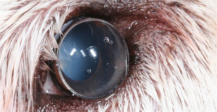
What Treatment Is There For Glaucoma In Dogs?
Treatment for glaucoma in dogs is lifelong once the disease has developed and includes eye drops, tablets and surgeries such as laser-treatment and the implantation of a variety of drainage shunts.
All treatments for glaucoma in dogs - even the eye drops - are costly and fail in the long term. Dogs with acute glaucoma are usually in extreme pain and the eyeball becomes swollen, the cornea blue and the white of the eye bright red.
DNA tests to identify glaucoma in dogs and those carrying the gene are starting to become available, but are currently only limited to a few breeds.
Christine Heinrich is the Director of the Eye Veterinary Clinic at Marlbrook, near Leominster in Herefordshire.